Skip to content

Free Printable Spinal Nerve Charts [Function & Diagram] PDF

    Understanding spinal anatomy and the network of nerves that transmit signals throughout the body is key for medical students and healthcare professionals. Learning the spinal nerves and their corresponding vertebral levels provides a foundation for diagnosing and treating spinal disorders and injuries. In this article, weโ€™ll explore how to read and utilize a spinal nerve chart to map innervation patterns.

    A downloadable spinal nerve chart PDF is included, listing each nerve, its origin vertebrae, and the region it enervates. With this chart, youโ€™ll have a handy visual aid to quickly reference the segments, roots, and branches of the elaborate spinal cord nervous system. Use the chart to memorize the arrangements and relationships between vertebrae, nerves, and extremities or as a practical guide for pathology analysis. Letโ€™s dive in to decoding the anatomy of spinal nerves.

    What Is A Spinal Nerve Chart?

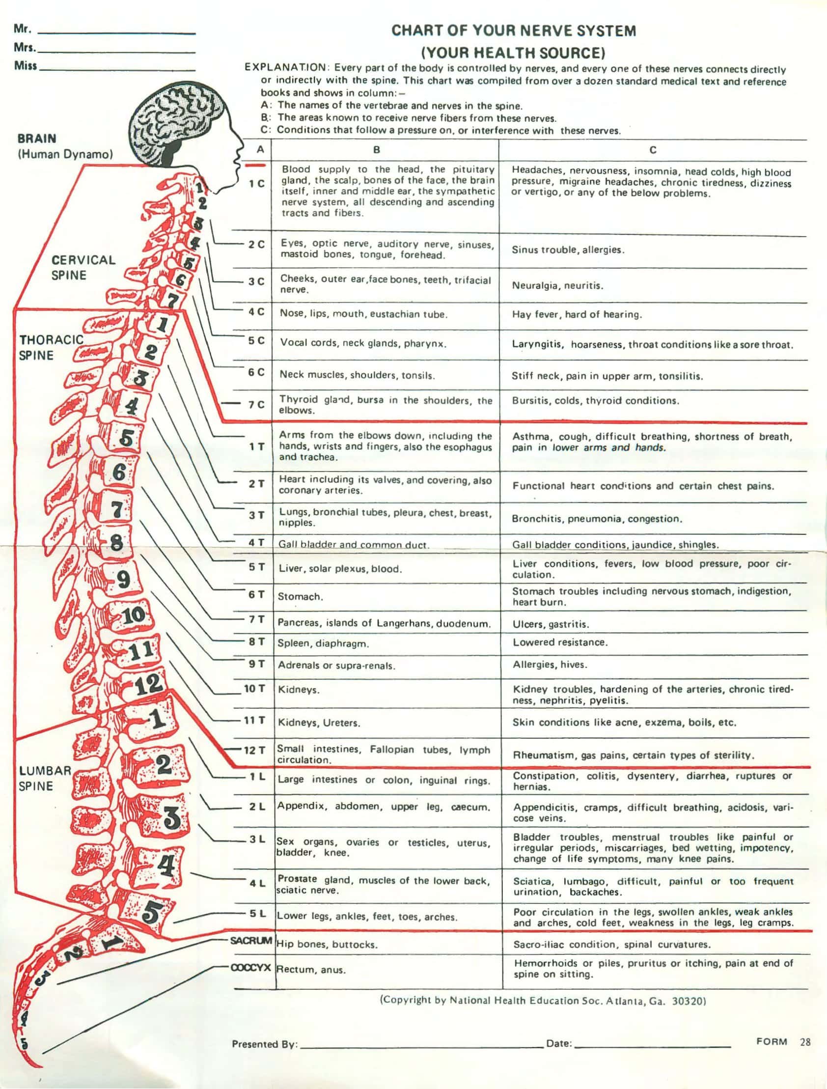
    Spinal Nerve Chart
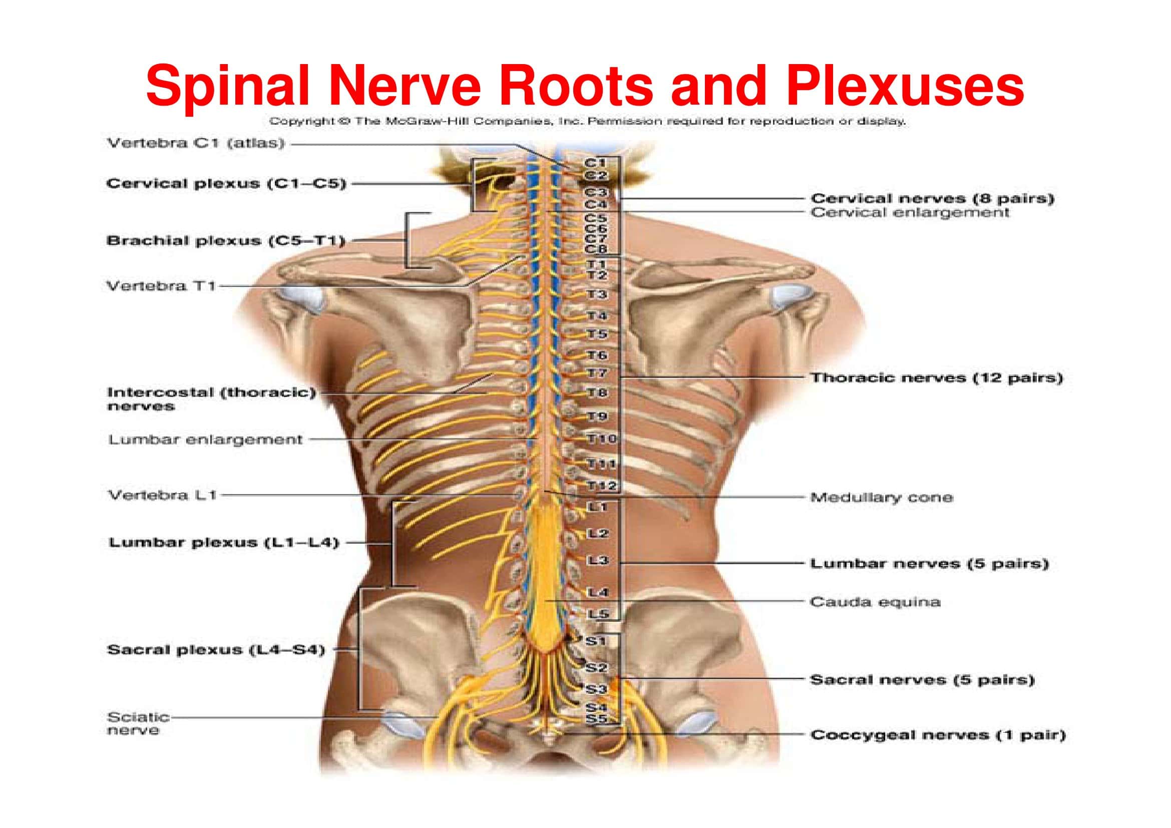
    Spinal Nerve Chart

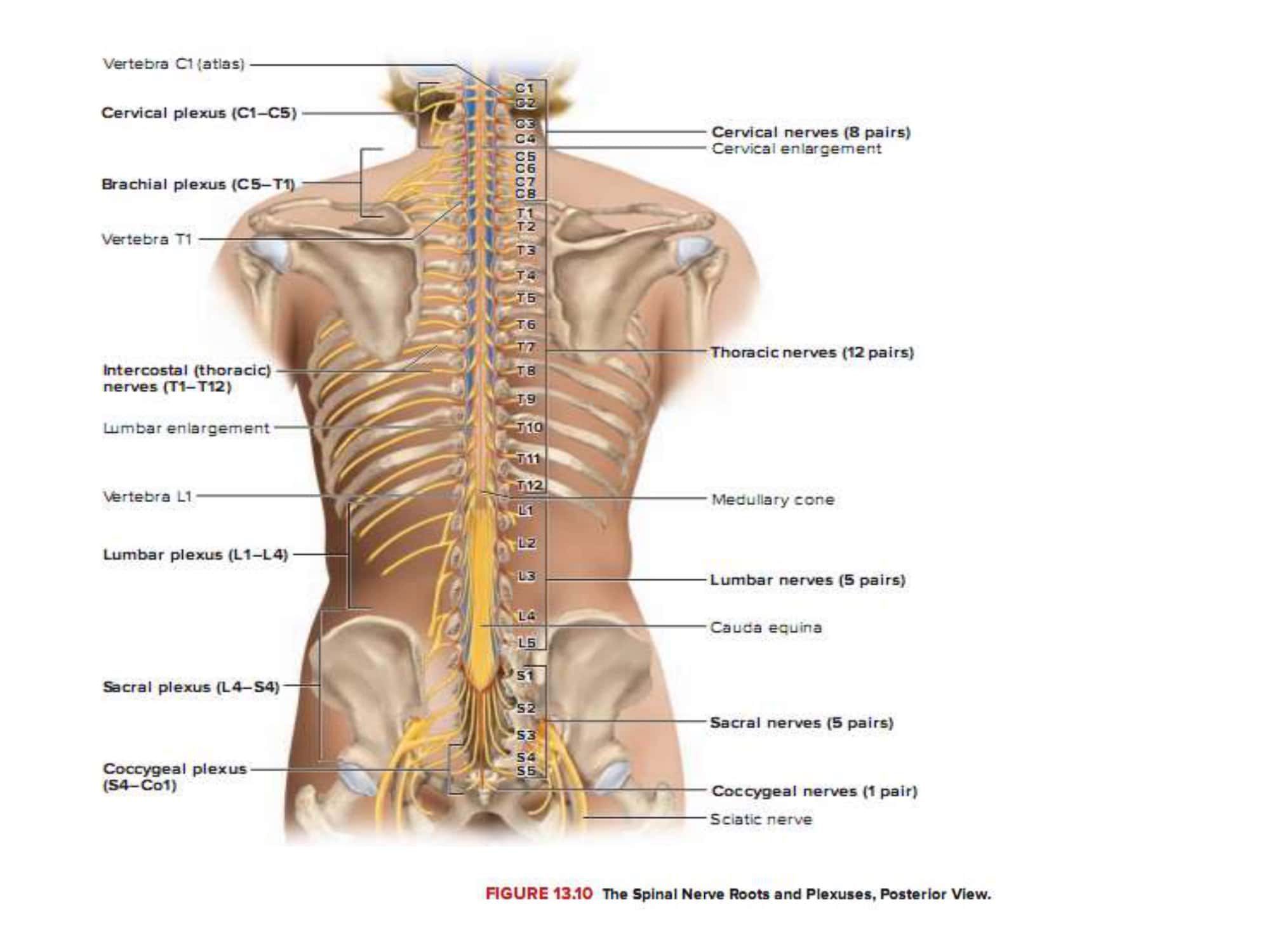
    A spinal nerve chart is a diagram that maps the spinal nerves branching from the vertebral column to the parts of the body they innervate. It depicts the cervical, thoracic, lumbar, and sacral sections of the spine, indicates the exit points of the nerve roots and how they combine into spinal nerves, and labels the sensory distribution of each nerve to the arms, torso, and legs.

    A spinal nerve chart provides a visual reference to help memorize the vertebral levels, sensory pathways, and motor functions of the network of nerves that transmit signals between the central nervous system and periphery. For medical professionals, it aids in pinpointing the origin of neurological issues based on affected dermatomes.

    Printable Spinal Nerve Chart

    The spinal nerve chart pdf is an anatomical diagram showing the nerves that exit from the spinal cord. It illustrates the cervical, thoracic, lumbar, and sacral regions of the spine and the associated spinal nerves. The chart depicts the intervertebral foramina between each vertebra where the spinal nerves exit. It labels the dorsal and ventral rami of each spinal nerve and shows how they innervate specific areas of the body.

    The pdf provides a visual reference for the 31 bilateral pairs of spinal nerves and their origins along the spinal cord. It presents the spinal nerves in sequential order, beginning with C1 exiting between the skull and C1 vertebra and descending to S5 between the L5 and S1 vertebrae. Key information is provided next to each spinal nerve including the spinal cord segment it arises from, its sensory distribution, and motor distribution.

    Overall, the spinal nerve chart pdf is an essential anatomical study aid. The clear and detailed illustrations with accompanying explanatory text outline the basic anatomy of the spinal cord, spinal nerves, nerve plexuses, and dermatomes. It serves as a useful tool for students looking to gain an understanding of how the central and peripheral nervous systems connect. The pdf format allows for convenient access anytime to recap this fundamental neuroanatomy content.

    Basic Anatomy of the Spinal Cord

    The spinal cord is a fundamental part of the central nervous system, transmitting neural signals between the brain and the rest of the body. Understanding the fundamental anatomy and organization of this intricate structure lays the groundwork for medical professionals to diagnose and treat spinal disorders. In this article, we will provide an overview of basic spinal cord anatomy, exploring key components like vertebrae, nerve roots, spinal segments, gray matter, white matter, and the integrating network of neuronal pathways.

    We will examine how the delicate spinal cord is protected within the vertebral column and its sectional organization into cervical, thoracic, lumbar, and sacral regions based on vertebral level. Additionally, we will look at the specialized posterior, anterior, and lateral spinal nerve roots that carry information between the cord and periphery. Gaining familiarity with core spinal cord anatomy and terminology provides the basis for appreciating its physiology and pathology. Letโ€™s review the foundational anatomy of this vital neurological structure.

    The Structure of the Spinal Cord:

    The spinal cord is a cylindrical structure of nervous tissue that extends from the medulla oblongata in the brainstem down to the lumbar region of the vertebral column. Enclosed within the vertebral column, it is continuous with the base of the brain and runs down to about the level of the second lumbar vertebra.

    The spinal cord is encased in the meninges and is cushioned by cerebrospinal fluid. It serves as the main pathway for transmitting neural signals between the brain and the rest of the body. The spinal cord has a butterfly or H-shaped area of gray matter at its center, surrounded by white matter. The gray matter contains neuron cell bodies, while the white matter consists of myelinated axons.

    Different Sections

    Cervical:
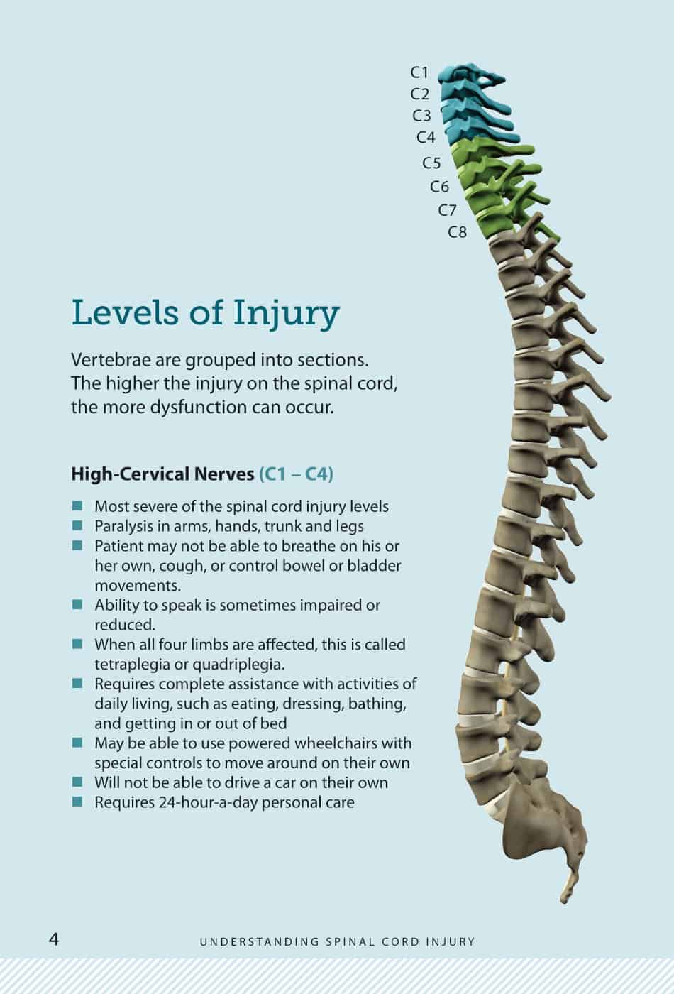
    The cervical section of the spinal cord is located in the neck and corresponds to the seven cervical vertebrae. This section contains eight pairs of cervical spinal nerves (C1 to C8). These nerves are responsible for sending signals to and from the neck, shoulders, arms, and upper chest. The most superior part, C1, is associated with the atlas vertebra and supports the head’s movements. Damage or injury to the cervical spinal cord can lead to quadriplegia, affecting both the arms and legs.

    Thoracic:
    The thoracic section of the spinal cord corresponds to the twelve thoracic vertebrae. It contains twelve pairs of thoracic spinal nerves (T1 to T12). These nerves are responsible for transmitting signals to and from the upper abdomen, mid-back, and chest. Unlike the cervical and lumbar enlargements, the thoracic region does not control limbs and therefore has a smaller amount of gray matter. The thoracic section primarily governs the muscles and structures of the chest and upper abdomen.

    Lumbar:
    The lumbar section of the spinal cord is associated with the five lumbar vertebrae. It contains five pairs of lumbar spinal nerves (L1 to L5). This section controls signals to and from the lower abdomen, hips, and legs. The lumbar section of the spinal cord is enlarged, similar to the cervical section, due to the need for innervation of the large muscles of the legs. Damage or injury to the lumbar spinal cord can result in paraplegia, affecting the lower limbs.

    Sacral and Coccygeal:
    The sacral part of the spinal cord corresponds to the five sacral vertebrae, which are fused in adults to form the sacrum. It contains five pairs of sacral spinal nerves (S1 to S5) that are responsible for transmitting signals to and from the lower parts of the body, including the buttocks, genitalia, and some parts of the leg and foot. The coccygeal region is the most terminal portion of the spinal cord and represents a vestige of the tail in our evolutionary ancestors. It typically has one pair of coccygeal spinal nerves. Together, the sacral and coccygeal nerves form the cauda equina, a bundle of nerves that looks like a horse’s tail, extending from the end of the spinal cord.

    Spinal Nerves: An In-depth Look

    Origin and Branching:
    Spinal nerves emerge as intricate structures that play a pivotal role in connecting the central nervous system to the rest of the body. Originating from the spinal cord, there are 31 pairs in total: 8 cervical, 12 thoracic, 5 lumbar, 5 sacral, and 1 coccygeal. Each of these nerves arises from the fusion of two primary roots โ€“ the dorsal (posterior) and the ventral (anterior) roots. Once these roots combine, they form a singular spinal nerve that exits the vertebral column through an intervertebral foramen. Shortly after emerging, each spinal nerve divides into several branches.

    The major branches include the dorsal ramus, which primarily serves the muscles and skin of the back, and the larger ventral ramus, which innervates the lateral and anterior parts of the trunk as well as the limbs. Additionally, there are meningeal branches that re-enter the vertebral canal to supply the vertebrae, vertebral ligaments, blood vessels of the spinal cord, and the meninges. Another significant branch is the rami communicantes, which connect the spinal nerves to the sympathetic trunk, a crucial part of the autonomic nervous system.

    The Dorsal and Ventral Roots:
    Diving deeper into the formation of the spinal nerves, it’s essential to understand the dorsal and ventral roots’ distinct roles and structures. The dorsal roots contain the sensory or afferent fibers. These fibers arise from sensory neurons whose cell bodies are located outside the spinal cord in specialized structures called dorsal root ganglia. These neurons pick up sensory information, such as touch, temperature, and pain from various parts of the body, and transmit this data to the spinal cord through the dorsal roots. On the flip side, the ventral roots comprise motor or efferent fibers.

    These fibers emanate from motor neurons located within the gray matter of the spinal cord. These motor neurons send impulses away from the spinal cord, instructing muscles to contract or glands to secrete. The unification of the sensory-laden dorsal roots with the motor-rich ventral roots within the spinal nerves ensures that both incoming sensory information and outgoing motor commands can be integrated and conveyed efficiently between the spinal cord and the distant regions of the body. This seamless integration and communication are foundational to our ability to interact with and respond to our environment.

    Spinal Nerves

    1. Cervical Spinal Nerves (C1-C8)

    • Anatomical Location: The cervical spinal nerves originate from the cervical region of the spinal cord, which is situated in the neck. There are eight pairs of cervical spinal nerves, labeled C1 to C8. They emerge above the vertebrae for which they are named; for example, the C1 nerve emerges above the first cervical vertebra (atlas). This pattern changes at the C7 vertebra, where the C8 nerve emerges below it.
    • C1 – Head and neck muscles
    • C2 – Eye and face movement
    • C3, C4 – Diaphragm, neck and shoulder movement
    • C5 – Shoulder and elbow motion
    • C6 – Wrist extension
    • C7 – Elbow flexion
    • C8 – Hand and finger control
    • Function: These nerves control signals to and from the neck, shoulders, arms, and hands. The first cervical nerve (C1) mainly supplies the muscles at the base of the skull, while C2 and C3 play roles in sensation for the upper neck and the back of the head. C4-C8 contribute to the formation of the brachial plexus which provides motor and sensory function to the arms and hands.
    • Common Injuries and Symptoms: Cervical nerve injuries can result from cervical disc herniation, whiplash, or other trauma. Symptoms can include neck pain, stiffness, and varying degrees of pain, numbness, or tingling radiating down the arm and into the hand. The specific location of these symptoms depends on which nerve is affected.

    2. Thoracic Spinal Nerves (T1-T12)

    • Anatomical Location: The thoracic spinal nerves emerge from the thoracic region of the spinal cord, which lies behind the rib cage. They exit the spinal column between the corresponding thoracic vertebrae.
    • T1-T6 – Intercostal and chest wall muscle control
    • T7-T9 – Muscles of the abdomen
    • T10-T12 – Muscles of the back
    • Function: These nerves primarily control the muscles and provide sensation to the torso or trunk. They play roles in the function of the rib cage, abdominal muscles, and some back muscles.
    • Common Injuries and Symptoms: Thoracic nerve injuries are less common due to the protection provided by the rib cage. However, injuries can still occur and result in pain around the rib cage or a band-like pain around the trunk, known as a “girdle” sensation. Herniated discs, compression fractures, and trauma are potential causes.

    3. Lumbar Spinal Nerves (L1-L5)

    • Anatomical Location: These nerves emerge from the lumbar region of the spinal cord, which is located in the lower back.
    • L1, L2 – Hip flexors and adductors
    • L3, L4 – Knee extension, ankle dorsiflexion
    • L5 – Foot intrinsic muscles
    • Function: Lumbar spinal nerves control muscles and provide sensation to the lower back, abdomen, buttocks, and parts of the legs.
    • Common Injuries and Symptoms: Lumbar nerve injuries often result from conditions like lumbar disc herniation or spinal stenosis. Symptoms can include low back pain, pain radiating down the legs (sciatica), numbness, tingling, and muscle weakness in the legs.

    4. Sacral Spinal Nerves (S1-S5)

    • Anatomical Location: These nerves originate from the sacral region of the spinal cord, located in the pelvis, behind the hip bone.
    • Function: Sacral spinal nerves are responsible for the motor and sensory functions of the buttocks, genitalia, thighs, and most of the lower legs and feet. They also play roles in bowel and bladder function.
    • S1-S3 – Hip, knee, ankle plantarflexion
    • S2-S4 – Pelvic floor, external anal sphincter
    • S5 – Foot intrinsic muscles
    • Common Injuries and Symptoms: Injuries can result from trauma or conditions like sacral disc herniation. Symptoms include pain, numbness, or tingling in the buttocks, backs of the thighs, and even into the feet. Bowel and bladder dysfunction can also occur in severe cases.

    5. Coccygeal Nerve (Co1)

    • Anatomical Location: This nerve emerges from the bottommost part of the spinal cord, the coccyx or tailbone.
    • Function: The coccygeal nerve provides sensation to the skin over the tailbone.
    • Common Injuries and Symptoms: Coccyx injuries, often termed as “tailbone injuries,” can result from direct trauma like falls. Symptoms include localized pain and tenderness at the base of the spine, particularly when sitting.

    How To Use The Spinal Nerve Chart:

    The spinal nerve chart, often referred to as a dermatome chart, illustrates the areas of sensory distribution for each spinal nerve. It helps to determine the possible level of spinal cord or nerve root involvement by noting the area of sensory disturbance, such as numbness or tingling. Here’s a detailed guide on how to use the spinal nerve chart:

    Understanding the Basics

    1. Know the Structure: Familiarize yourself with the layout of the chart. It should show a front and back view of a human body, with various sections shaded or colored differently, each labeled with a specific nerve’s designation (e.g., C5, T3, L2).
    2. Dermatomes: Each spinal nerve provides sensation to a specific region of the skin called a dermatome. Understand that the overlap between dermatomes means that a single patch of skin often receives input from multiple spinal nerves.

    Using the Chart

    1. Identify the Affected Area: Determine the area of the body where there’s numbness, tingling, pain, or other abnormal sensations.
    2. Match to the Chart: Locate this area on the dermatome chart to identify which spinal nerve might be affected. For instance, if someone complains of numbness on the outer thigh, you’d look for this area on the chart and see it corresponds to the L2 dermatome.
    3. Consider Overlaps: Remember that due to overlaps, a complete loss of sensation in a dermatome usually requires damage to more than one spinal nerve. If only one nerve is damaged, there might still be some sensation due to the overlap from neighboring nerves.
    4. Compare Bilaterally: Compare the sensation on both sides of the body. If one side has abnormal sensation while the other doesn’t, it can indicate a unilateral issue at a specific spinal level.

    Additional Tips for Professionals

    1. Clinical Examination: For medical professionals, using a pinprick or soft cotton wool can help determine areas of reduced or heightened sensation. Ask the patient to compare the sensation in the suspected area to a nearby ‘normal’ area.
    2. Motor Function: The chart might also show the major muscles or movements controlled by each nerve. Check for weakness or abnormalities in these movements to further pinpoint the affected nerve.
    3. Reflex Testing: Some charts also include the reflexes associated with specific spinal levels. For instance, the patellar reflex is linked to the L3 and L4 spinal levels.
    4. Be Comprehensive: Remember, a single symptom or test does not provide a definitive diagnosis. Other conditions like peripheral neuropathies or systemic diseases can also cause sensory changes. Use the chart as a tool within a comprehensive clinical assessment.
    5. Stay Updated: Dermatome distributions can slightly vary among different charts. Stick to reputable and updated sources, and understand that individual variations can occur.

    Using the Chart for Treatment

    1. Localization: Once you’ve identified the potential affected nerve or spinal segment, you can target treatments. This could involve therapies, exercises, or interventions specific to that level.
    2. Monitoring: Over time, as treatment progresses, use the chart to monitor changes in sensation or function. Any improvement or worsening can guide adjustments in treatment.

    Modern Imaging Techniques

    Advancements:

    1. Magnetic Resonance Imaging (MRI): Over the years, MRI technology has witnessed tremendous improvements. High-resolution MRI now provides clearer images of soft tissues, including intervertebral discs, spinal cord, and nerve roots. This has improved the accuracy of diagnoses for conditions like disc herniation and spinal cord pathologies.
    2. Functional MRI (fMRI): Unlike traditional MRI, fMRI captures rapid dynamic changes and can map the active parts of the brain or spinal cord by measuring changes associated with blood flow. This helps in understanding the functional aspects of the spine and brain.
    3. Diffusion Tensor Imaging (DTI): A variant of MRI, DTI maps the pathways of white matter tracts in the nervous system. This provides information about nerve pathways, helping in planning surgeries or understanding complex nerve injuries.

    Future Prospects:

    1. Portable Imaging Devices: With technology becoming compact, we can foresee the development of portable spine imaging devices. This would greatly aid in point-of-care diagnostics and treatments.
    2. AI Integration: Artificial Intelligence is steadily becoming a part of radiological imaging, assisting in rapid, precise image interpretation, and disease detection.

    Digital and Interactive Spinal Nerve Charts

    Advancements:

    1. Digital Accessibility: Traditional paper charts have given way to digital charts that are easily accessible on tablets, computers, and even smartphones. This provides healthcare providers with instantaneous access to these charts, aiding in faster clinical decision-making.
    2. Interactive Features: Modern digital spinal nerve charts come equipped with interactive features. Clinicians can zoom in on specific areas, overlay different anatomical structures, and sometimes even get 3D views.
    3. Integrated Patient Data: Some advanced systems allow integration of patient-specific data with the charts. For example, an MRI of a patient can be overlayed onto the standard spinal nerve chart, giving a personalized view.
    4. Augmented Reality (AR) & Virtual Reality (VR): AR and VR technologies are starting to find their place in spinal nerve chart interpretations. They allow for immersive 3D models, where clinicians can “walk through” the anatomical structures or even simulate surgical procedures.

    Future Prospects:

    1. Personalized Digital Charts: In the future, digital spinal nerve charts might be routinely integrated with patients’ own imaging data, providing a personalized anatomy chart for every individual.
    2. Remote Sharing and Consultations: With the advancement of telemedicine, these interactive charts can be shared in real-time with specialists anywhere in the world, aiding in remote consultations and collaborative treatments.
    3. Educational Use: The interactive features, combined with AR and VR, can revolutionize how medical students learn about the spine and nervous system. Instead of static images, they can interact with dynamic, 3D models, enhancing understanding and retention.

    Conclusion

    Navigating the intricate anatomy of the spinal nerves from roots to terminations is essential, yet complex, knowledge in medicine. This article has explored utilizing spinal nerve charts to visualize the organization and connections of this neural highway. With the included downloadable PDF reference, you can now easily map the network of cervical, thoracic, lumbar, sacral and coccygeal nerves to their corresponding vertebrae and dermatomes.

    Use the chart to identify spinal levels for sensory pathways or motor functions, locate sources of radiculopathies, and pinpoint which muscles or dermatomes a nerve supplies. Keep this guide close-by in your studies or clinic for quick reference to spinal nerve innervations, symptoms and pathology. Whether you are actively learning the concepts or need a refresher, this chart is an invaluable tool for clarifying the anatomy. Master the critical anatomy of the spinal cord nerves with this free, handy reference.

    FAQs

    Why is the spinal nerve chart important?

    The chart is crucial for clinicians to quickly localize potential nerve or spinal cord issues based on a patient’s reported area of numbness, tingling, or pain. It aids in accurate diagnosis, treatment planning, and patient education.

    How many spinal nerves are there?

    There are 31 pairs of spinal nerves: 8 cervical, 12 thoracic, 5 lumbar, 5 sacral, and 1 coccygeal.

    What is a dermatome?

    A dermatome is a specific area of the skin that is innervated by a single spinal nerve. If there’s a problem with a particular spinal nerve, it can result in sensory disturbances in its corresponding dermatome.

    How do spinal nerve charts help in diagnosis?

    By comparing a patient’s reported sensory symptoms with the dermatome chart, clinicians can narrow down which spinal nerve(s) or segment(s) of the spinal cord might be affected. This aids in further diagnostic testing and treatment.

    Are there overlaps in dermatomes?

    Yes, dermatomes often overlap, meaning a particular area of the skin might receive sensations from multiple spinal nerves. Therefore, a complete loss of sensation in a dermatomal area often requires damage to more than one spinal nerve.

    Can a spinal nerve chart help in understanding back pain?

    Yes, while not all back pain is directly related to spinal nerve issues, the chart can help pinpoint potential nerve involvement. For example, pain radiating down the leg following a specific pathway might indicate sciatic nerve involvement, which is related to certain lumbar nerve roots.

    Click to rate this post!
    [Total: 3 Average: 5]
    Betina Jessen

    Betina Jessen

    Leave a Reply

    Your email address will not be published. Required fields are marked *