The human mouth, a complex and fascinating organ, is much more than a conduit for nutrition—it is an intricate system where each component plays a crucial role. One of the most vital parts are our teeth, stalwart soldiers of digestion that prepare food for our stomach.
Yet, despite their daily importance, many of us aren’t aware of the vast diversity and unique roles of different types of teeth. In this article, we delve deep into the world of dental anatomy, exploring the interesting and informative tooth chart. From incisors to molars, we’ll decode the mysteries of each tooth’s shape, location, and function, offering you a comprehensive guide to understanding this fundamental aspect of human biology. So get ready to embark on a captivating journey, one that will forever change your perception of a smile.
Table of Contents
What is a Tooth Chart ?
![Free Printable Tooth Chart Templates – [Dental Chart] Teeth Numbers PDF 1 Tooth Chart](https://www.typecalendar.com/wp-content/uploads/2023/06/Tooth-Chart-1024x576.jpg 1024w, https://www.typecalendar.com/wp-content/uploads/2023/06/Tooth-Chart-300x169.jpg 300w, https://www.typecalendar.com/wp-content/uploads/2023/06/Tooth-Chart-768x432.jpg 768w, https://www.typecalendar.com/wp-content/uploads/2023/06/Tooth-Chart-1536x864.jpg 1536w, https://www.typecalendar.com/wp-content/uploads/2023/06/Tooth-Chart-1200x675.jpg 1200w, https://www.typecalendar.com/wp-content/uploads/2023/06/Tooth-Chart.jpg 1920w)
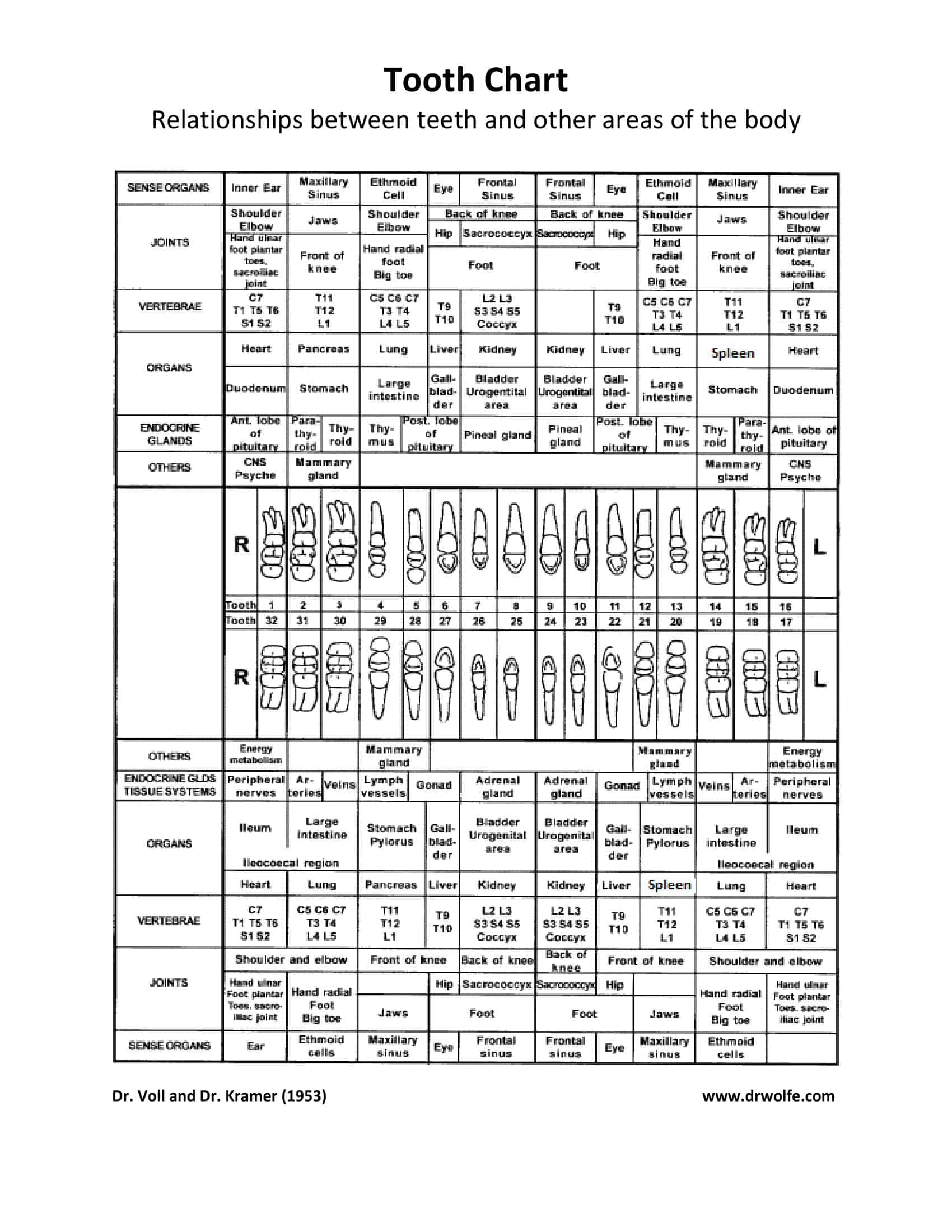
A tooth chart, also known as a dental chart, is a graphical representation of the teeth present in the mouth. It serves as a visual guide to the types, positions, and numbers of teeth in both children (primary or “baby” teeth) and adults (permanent teeth).
In its most basic form, a tooth chart illustrates the location and name of each tooth, including incisors, canines, premolars, and molars. It’s typically divided into four quadrants (upper left, upper right, lower left, and lower right) to correspond to the layout of the human mouth. Each tooth is identified by a specific number or letter, following either the Universal Numbering System (used primarily in the United States) or the FDI World Dental Federation notation (international standard).
In more detailed versions, a tooth chart can also provide information about the structure of each tooth, including layers such as the enamel, dentin, pulp, and the surrounding structures like gums and bone.
Tooth Chart Templates
Importance of Tooth Charts in Dentistry
Tooth charts play an instrumental role in dentistry, helping to streamline communication, diagnosis, treatment planning, and patient education. Below, we explore the manifold importance of tooth charts in the world of dental health:
1. Standardized Communication
Tooth charts provide a universal language for dental professionals. Regardless of geographic location, dentists can utilize the standardized numbering or lettering systems (Universal or FDI World Dental Federation notation) to identify specific teeth quickly and accurately. This uniformity facilitates clear communication between different practitioners or specialists involved in a patient’s care, ensuring everyone is on the same page.
2. Diagnosis & Treatment Planning
Tooth charts are indispensable in diagnosing dental issues and crafting treatment plans. By marking areas of concern on a tooth chart, such as cavities, tooth decay, gum disease, or tooth loss, dentists can visualize the scope of the problem and develop an appropriate treatment strategy. Tooth charts can also be used to track the progression of these conditions over time.
3. Record Keeping
Detailed dental charts form an integral part of a patient’s dental record. They provide an at-a-glance summary of a patient’s dental history, including prior procedures, tooth extractions, implants, or orthodontic treatments. This historical data is invaluable for long-term patient management and can inform future treatment decisions.
4. Patient Education
Tooth charts are excellent visual aids for patient education. Dentists can use these charts to explain dental conditions, procedures, and oral hygiene practices in an easily understandable way. For example, a dentist might use a tooth chart to show a patient where a cavity has developed and how a filling or root canal procedure would address the problem.
5. Tracking Dental Development
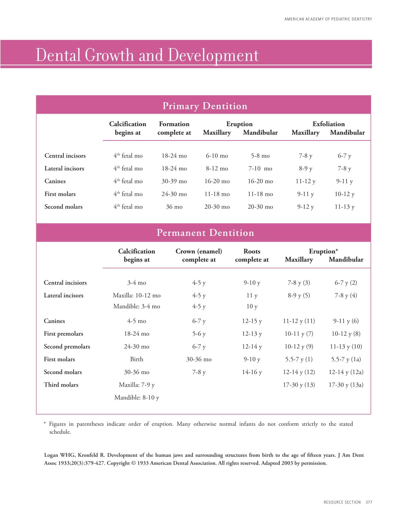
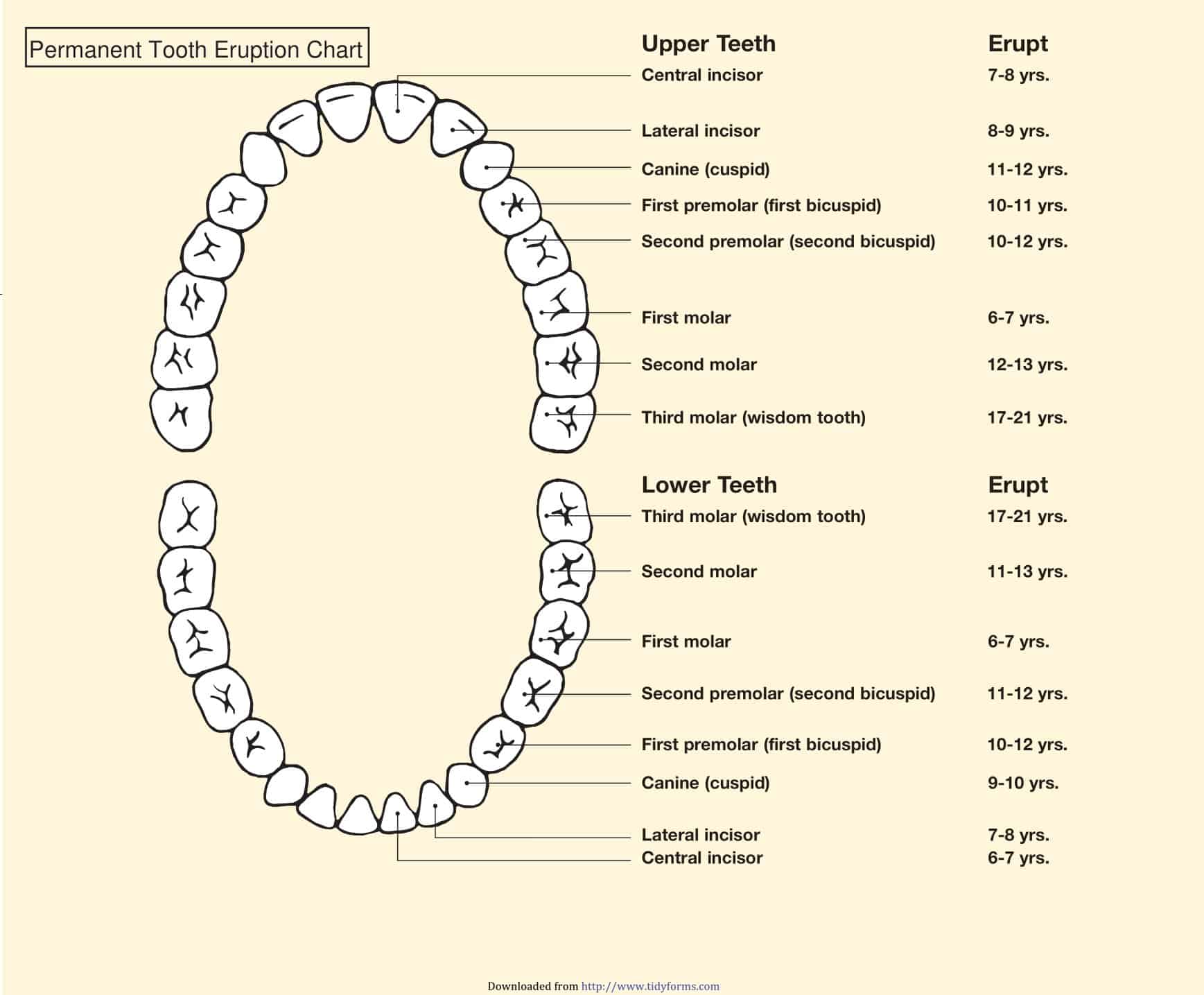
For pediatric dentistry, tooth charts are essential for tracking the development and eruption of primary (baby) and permanent (adult) teeth. Any deviations from the typical pattern can be noted and monitored, helping to identify potential issues such as impacted teeth, early or late tooth loss, or the need for orthodontic intervention.
6. Preoperative and Postoperative Evaluation
Tooth charts can be used to plan complex dental procedures, such as implant placement, root canal treatment, or orthodontic work. They also serve as a reference during postoperative assessments, helping to evaluate the success of the treatment and plan for any necessary follow-up care.
Types of Tooth Charts
Tooth charts come in various types, each designed for a specific purpose, context, or age group. Below, we will explore some of the main types of tooth charts and their unique attributes.
1. Universal Tooth Chart:
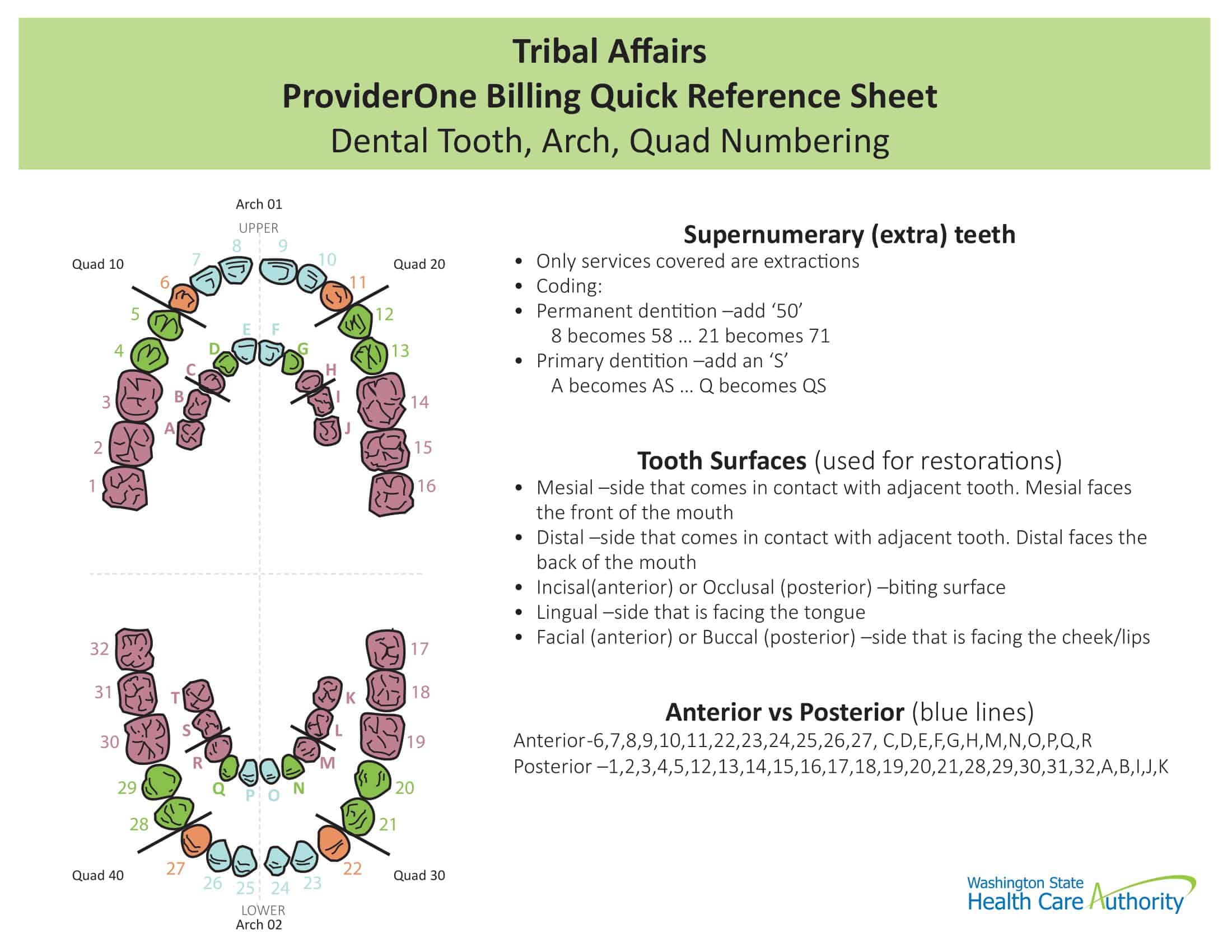
This is the most commonly used tooth chart in the United States. It numbers the teeth from 1-32, with number 1 being the right upper third molar (wisdom tooth) and number 32 being the right lower third molar. This chart is divided into four quadrants, with the upper right quadrant containing teeth 1-8, upper left containing teeth 9-16, lower left containing teeth 17-24, and lower right containing teeth 25-32. This is a simple and straightforward system but isn’t widely used outside the U.S.
2. Palmer Notation Method:
The Palmer notation method uses a combination of numbers (1-8) and symbols to identify teeth. The teeth in each quadrant are numbered from the central incisor (number 1) to the third molar (number 8). A unique symbol (a right angle) is used to designate each of the four quadrants. The symbols point in different directions: upwards for upper quadrants and downwards for lower quadrants, with the corner of the angle on the side of the midline. This system is often used in orthodontics.
3. FDI World Dental Federation Notation (ISO System):
The FDI system is an international standard for tooth numbering. It uses a two-digit number, where the first digit represents the quadrant and the second digit represents the tooth within that quadrant. Quadrants are numbered as follows: upper right (1), upper left (2), lower left (3), and lower right (4). Within each quadrant, teeth are numbered from the central incisor (1) to the third molar (8). So, for example, the upper right first molar is 16, while the lower left canine is 33. This system is widely used globally due to its simplicity and precision.
4. Primary Tooth Chart:
A primary tooth chart is specifically designed to document and track the development and eruption of primary (baby) teeth. This chart typically includes 20 teeth: four incisors, two canines, and four molars in each of the two arches (upper and lower). The Universal and FDI systems both have versions for primary teeth. In the Universal system, primary teeth are designated by letters A through T. In the FDI system, primary teeth are represented with a quadrant number (5 for upper right, 6 for upper left, 7 for lower left, and 8 for lower right) and a tooth number (1-5).
5. Orthodontic Tooth Chart:
Orthodontic tooth charts are specialized charts used for planning and tracking orthodontic treatments, such as braces or aligners. They often include additional details like the angulation and rotation of teeth, the presence of spaces or crowding, and notes on occlusion (bite).
6. Periodontal Chart:
This type of chart is used to document the health of the gums and supporting tooth structures. It includes spaces to record measurements of gum pocket depth, gingival recession, areas of bleeding or inflammation, and bone loss. It’s a critical tool for diagnosing and managing periodontal (gum) disease.
7. Dental Implant Chart:
Dental implant charts record important information about placed implants, such as their location, size, angle, and the brand or type of implant used. They also provide space for notes on the health of the surrounding tissues and the outcome of the implant procedure.
Anatomy of a Tooth
The human tooth is a complex structure that extends beyond the white, visible portion we see when we smile. Each tooth is made up of several layers, with each one playing a specific role. In addition to the layers, each tooth also has different surfaces with distinct names. Below we will break down these elements in more detail.
A. Tooth Structure and Function
Each tooth consists of two main parts: the crown and the root. The crown is the visible part of the tooth, while the root is embedded in the jawbone, stabilizing the tooth in its socket. These two parts are divided into several layers, each with its own unique structure and function.
1. Enamel: The enamel forms the outermost layer of the crown. It’s the hardest substance in the human body and is composed mostly of mineral, specifically hydroxyapatite crystals. The primary function of enamel is to protect the tooth from physical and chemical damage, especially during biting and chewing.
2. Dentin: Dentin lies directly beneath the enamel and forms the bulk of the tooth structure. It’s not as hard as enamel but is still quite mineralized. Dentin serves as a protective layer and also transmits signals from the exterior of the tooth to the nerves inside, causing sensitivity or pain when exposed due to cavities or tooth wear.
3. Pulp: The pulp is the innermost layer of the tooth and contains nerves, blood vessels, and connective tissue. The pulp supplies nutrients to the tooth and produces dentin. It also transmits sensory information (like pain or temperature) to the brain.
4. Cementum: Cementum is a bone-like substance covering the tooth root. It helps attach the tooth to the jawbone via the periodontal ligament.
5. Periodontal Ligament: This specialized connective tissue connects the tooth’s cementum with the alveolar bone of the jaw, helping to hold the tooth in place and absorb shocks from biting and chewing.
B. Tooth Surfaces and Nomenclature
Each tooth in the mouth has five surfaces, each with a unique name. These names help dentists precisely locate and describe areas of decay, damage, or treatment.
1. Occlusal/Biting Surface: This is the top surface of premolars and molars – the teeth at the back of the mouth. For incisors and canines, it’s referred to as the “incisal” edge.
2. Mesial Surface: The mesial surface of a tooth is the side closest to the midline of the face (the line that divides the face into two equal halves).
3. Distal Surface: The distal surface is the side of the tooth farthest from the midline.
4. Buccal/Facial Surface: The buccal surface (also known as the facial surface for front teeth) is the side of the tooth facing the cheeks or lips. For the front teeth, this surface can also be called “labial.”
5. Lingual/Palatal Surface: The lingual surface is the side of the tooth facing the tongue. For the upper teeth, this surface can also be called “palatal,” as it faces the palate (roof of the mouth).
Understanding the anatomy of a tooth, its structure, and the nomenclature of tooth surfaces is key to understanding oral health and disease. It’s also essential for communicating effectively with dental professionals.
Teeth Numbers and Names
Here’s a detailed breakdown of teeth numbers and names in both the Universal Numbering System (used mainly in the U.S.) and the FDI World Dental Federation notation (used internationally).
Universal Numbering System
Upper Jaw (Right to Left from the patient’s perspective):
- Number 1: Third Molar, often called the right upper wisdom tooth
- Number 2: Second Molar, often called the right upper 12-year molar
- Number 3: First Molar, often called the right upper 6-year molar
- Number 4: Second Bicuspid, also known as the right upper second premolar
- Number 5: First Bicuspid, also known as the right upper first premolar
- Number 6: Canine (Cuspid), also known as the right upper canine or eye tooth
- Number 7: Lateral Incisor, often called the right upper lateral incisor
- Number 8: Central Incisor, often called the right upper central incisor
- Number 9: Central Incisor, often called the left upper central incisor
- Number 10: Lateral Incisor, often called the left upper lateral incisor
- Number 11: Canine (Cuspid), also known as the left upper canine or eye tooth
- Number 12: First Bicuspid, also known as the left upper first premolar
- Number 13: Second Bicuspid, also known as the left upper second premolar
- Number 14: First Molar, often called the left upper 6-year molar
- Number 15: Second Molar, often called the left upper 12-year molar
- Number 16: Third Molar, often called the left upper wisdom tooth
Lower Jaw (Left to Right from the patient’s perspective):
- Number 17: Third Molar, often called the left lower wisdom tooth
- Number 18: Second Molar, often called the left lower 12-year molar
- Number 19: First Molar, often called the left lower 6-year molar
- Number 20: Second Bicuspid, also known as the left lower second premolar
- Number 21: First Bicuspid, also known as the left lower first premolar
- Number 22: Canine (Cuspid), also known as the left lower canine or eye tooth
- Number 23: Lateral Incisor, often called the left lower lateral incisor
- Number 24: Central Incisor, often called the left lower central incisor
- Number 25: Central Incisor, often called the right lower central incisor
- Number 26: Lateral Incisor, often called the right lower lateral incisor
- Number 27: Canine (Cuspid), also known as the right lower canine or eye tooth
- Number 28: First Bicuspid, also known as the right lower first premolar
- Number 29: Second Bicuspid, also known as the right lower second premolar
- Number 30: First Molar, often called the right lower 6-year molar
- Number 31: Second Molar, often called the right lower 12-year molar
- Number 32: Third Molar, often called the right lower wisdom tooth
Note: Teeth numbers 1-16 are in the upper arch of the mouth while numbers 17-32 are in the lower arch. The right and left sides are determined from the patient’s view.
In the Universal System, primary (baby) teeth are designated by letters A through T. Starting with the upper right second molar as A, the letters continue along the upper arch to the upper left second molar, which is J. The designation continues on the lower arch starting with the lower left second molar as K and ending with the lower right second molar as T.
This detailed breakdown should help you understand the layout and naming conventions of the teeth in the Universal Numbering System.
Tooth Charting Techniques
Tooth charting is a critical practice in dentistry that helps keep track of the health status of each tooth in a patient’s mouth. This methodical process involves the recording of information related to the structure, pathology, restorations, and relevant procedures performed on every tooth. There are several techniques used in tooth charting, ranging from traditional manual methods to innovative digital systems and advancements in charting technology.
A. Manual Charting Methods
Manual tooth charting is the traditional method and was the go-to technique before the advent of digital systems. Here’s how it typically works:
- Use of Dental Mirrors and Probes: Dentists use dental mirrors for visual inspection and dental probes for tactile examination. This allows them to identify and record issues such as caries (cavities), restorations (fillings, crowns), missing teeth, and other conditions.
- Recording Findings: The findings are usually noted on a paper tooth chart that resembles the layout of the teeth in the mouth. Dentists use a system of symbols and abbreviations to mark the status of each tooth. For example, a circle might indicate a filling, an “X” might represent an extracted tooth, and so on.
- Updating Records: The manual chart must be updated during each visit to reflect any changes since the last appointment.
While manual charting is cost-effective and requires no special equipment, it can be time-consuming and prone to human error. Additionally, paper charts can take up a significant amount of physical storage space and may degrade over time.
B. Digital Charting Systems
Digital tooth charting has become increasingly popular due to its efficiency, accuracy, and ease of use. Digital systems allow dental professionals to chart findings using a computer or a tablet. The advantages of digital charting include:
- Efficiency and Accuracy: Digital charting is generally faster than manual methods. It can provide clearer, more precise representations of teeth and their conditions. Additionally, the risk of human error is reduced.
- Integration with Other Digital Systems: Digital charts can be integrated with other systems, such as digital radiographs and intraoral cameras. This allows for a comprehensive view of the patient’s oral health in one place.
- Ease of Updating and Retrieving Records: Digital records are easy to update, and historical data can be accessed and compared at the click of a button.
Despite these advantages, implementing a digital charting system can be expensive and requires training to use effectively. Also, issues like technical glitches and data security need to be managed carefully.
C. Advancements in Tooth Charting Technology
As dental technology continues to evolve, new methods are emerging that improve the ease, accuracy, and efficiency of tooth charting.
- 3D Dental Imaging: Advanced imaging technologies such as Cone Beam Computed Tomography (CBCT) provide 3D visualizations of the teeth and surrounding structures. This allows for more accurate charting, especially for complex conditions and treatments.
- Artificial Intelligence (AI): AI algorithms can assist in automating the tooth charting process. For example, AI can analyze radiographs or intraoral images to detect and chart cavities, restorations, and other features.
- Virtual and Augmented Reality: Virtual Reality (VR) and Augmented Reality (AR) technologies can provide interactive 3D models for charting. These technologies can provide more intuitive and detailed visualizations compared to traditional 2D charts.
Applications of Tooth Charts
Tooth charts are a crucial tool in dentistry, with diverse applications ranging from clinical care to research and education. They provide a visual representation of a patient’s oral health status and can be used to plan treatments, communicate information, track changes over time, and conduct research. Below, we explore three primary areas where tooth charts are routinely used.
1. Dental Examinations and Diagnoses
Tooth charts are invaluable during dental examinations and diagnoses. They provide a structured method for recording the status of each tooth and related structures.
- Assessment: During an oral examination, the dentist systematically inspects each tooth and notes any findings on the tooth chart. This might include observations about the tooth’s condition, such as the presence of caries, fractures, restorations, or signs of periodontal disease.
- Diagnosis: The detailed information captured on a tooth chart aids in diagnosing oral health problems. For instance, a chart might reveal a pattern of decay suggestive of a particular dietary habit, or the location of periodontal pockets might point towards specific periodontal diseases.
2. Treatment Planning and Documentation
Tooth charts play a central role in treatment planning and documentation.
- Treatment Planning: Once a diagnosis has been made, the dentist uses the tooth chart as a blueprint for treatment planning. The chart helps identify which teeth need intervention and the type of treatment required, whether it’s a filling, root canal therapy, extraction, or periodontal treatment.
- Documentation: Tooth charts serve as a vital record of the treatments performed. If a tooth has been filled or extracted, this is marked on the chart. This record is useful for both the dentist and the patient as it provides a history of dental work completed, aids in future treatment planning, and tracks the progression of oral health over time.
3. Dental Research and Education
Tooth charts also have significant applications in dental research and education.
- Research: In dental research, tooth charts can provide data on patterns of oral diseases, the efficacy of treatments, and trends over time. For instance, researchers can analyze historical tooth charts to study the prevalence of dental caries in certain populations.
- Education: In dental education, tooth charts are used to teach dental students about the anatomy and pathology of teeth. Students learn how to chart teeth and interpret tooth charts as part of their clinical training. Tooth charts are also used in patient education, helping dentists explain diagnoses and treatments to their patients visually.
In summary, tooth charts are a versatile tool in dentistry, underpinning everything from patient care to research and education. Their use enhances communication, aids in diagnosis and treatment planning, provides a historical record, and facilitates research and learning.
Conclusion
Embracing the complexities of the dental landscape, tooth charts serve as an integral resource for dental professionals, researchers, and educators alike. They meticulously document each tooth’s condition, aid in accurate diagnoses, and provide a strategic roadmap for treatment plans. Equally important, they stand as a reliable source of historical data for ongoing research and as a visual aid for educating aspiring dentists and patients.
As digital innovations and advanced technologies continue to evolve, tooth charts are also poised for transformation, offering even more precision and efficiency. Ultimately, the essence of the tooth chart lies in its ability to reflect our oral health, providing insights that guide us towards improved dental care and a healthier future.
FAQs
How are tooth charts used in dental research and education?
In dental research, tooth charts can provide data on patterns of oral diseases, the efficacy of treatments, and trends over time. In dental education, tooth charts are used to teach dental students about the anatomy and pathology of teeth. Students learn how to chart teeth and interpret tooth charts as part of their clinical training.
What is tooth charting in a dental examination?
Tooth charting in a dental examination involves the dentist systematically inspecting each tooth, noting its condition, and recording this information on the tooth chart. This might include the presence of caries (cavities), restorations (fillings, crowns), periodontal disease, or any other noteworthy observations.
What does it mean if a tooth is marked with an ‘X’ on a tooth chart?
In many charting systems, a tooth marked with an ‘X’ indicates that the tooth has been extracted. However, the meaning can vary depending on the system being used and the dentist’s notation style, so it’s best to ask your dental professional for clarification.
Can tooth charts be used to identify human remains?
Yes, tooth charts can play a vital role in forensic odontology, which includes the identification of human remains. Teeth are remarkably durable and can often survive when other means of identification are not possible. A comparison of dental records, including tooth charts, with the teeth of unidentified remains can help establish the identity of a deceased individual.


![Free Printable Pie Chart Templates [Excel, PDF, Word] Maker 2 Pie Chart](https://www.typecalendar.com/wp-content/uploads/2023/06/Pie-Chart-150x150.jpg 150w, https://www.typecalendar.com/wp-content/uploads/2023/06/Pie-Chart-1200x1200.jpg 1200w)
![Free Printable Reward Chart Templates [Word, PDF] Teachers 3 Reward Chart](https://www.typecalendar.com/wp-content/uploads/2023/03/Reward-Chart-150x150.jpg 150w, https://www.typecalendar.com/wp-content/uploads/2023/03/Reward-Chart-1200x1200.jpg 1200w)
![Free Printable Behavior Chart Templates [PDF, Word, Excel] 4 Behavior Chart](https://www.typecalendar.com/wp-content/uploads/2023/02/Behavior-Chart-150x150.jpg 150w, https://www.typecalendar.com/wp-content/uploads/2023/02/Behavior-Chart-1200x1200.jpg 1200w)
